
Manual Patch Clamp Ion Channel Screening Services
Manual patch-clamp
Manual patch-clamp (MPC) was developed by Erwin Neher and Bert Sackman in 1976 and subsequently awarded a nobel prize in 1991. Facilitating recording of electrical events from single cells in real-time, MPC provides a powerful and versatile technique which has revolutionised our understanding of cardio- and neurophysiology.
Cultured cells or ex vivo tissue preparations are perfused in a recording chamber attached to a microscope, allowing the user to visually select cells of interest. Glass microelectrodes are positioned at the cell membrane and suction is applied to form an electrically tight seal. Summated electrical events can be recorded in cell-attached or whole-cell configurations (Figure 1), or alternatively small patches of membrane can be isolated and single ion-channel events recorded (outside- or inside-out configurations).
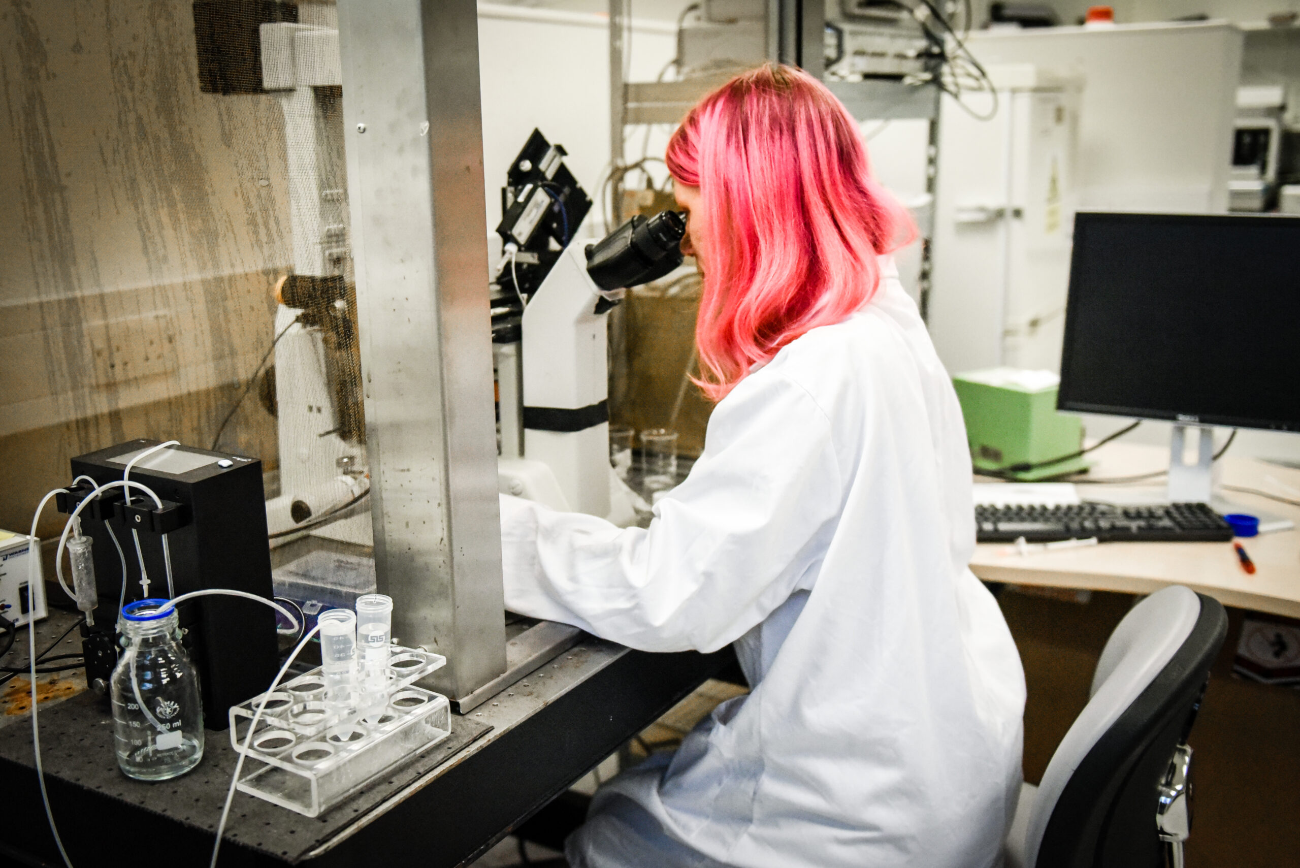
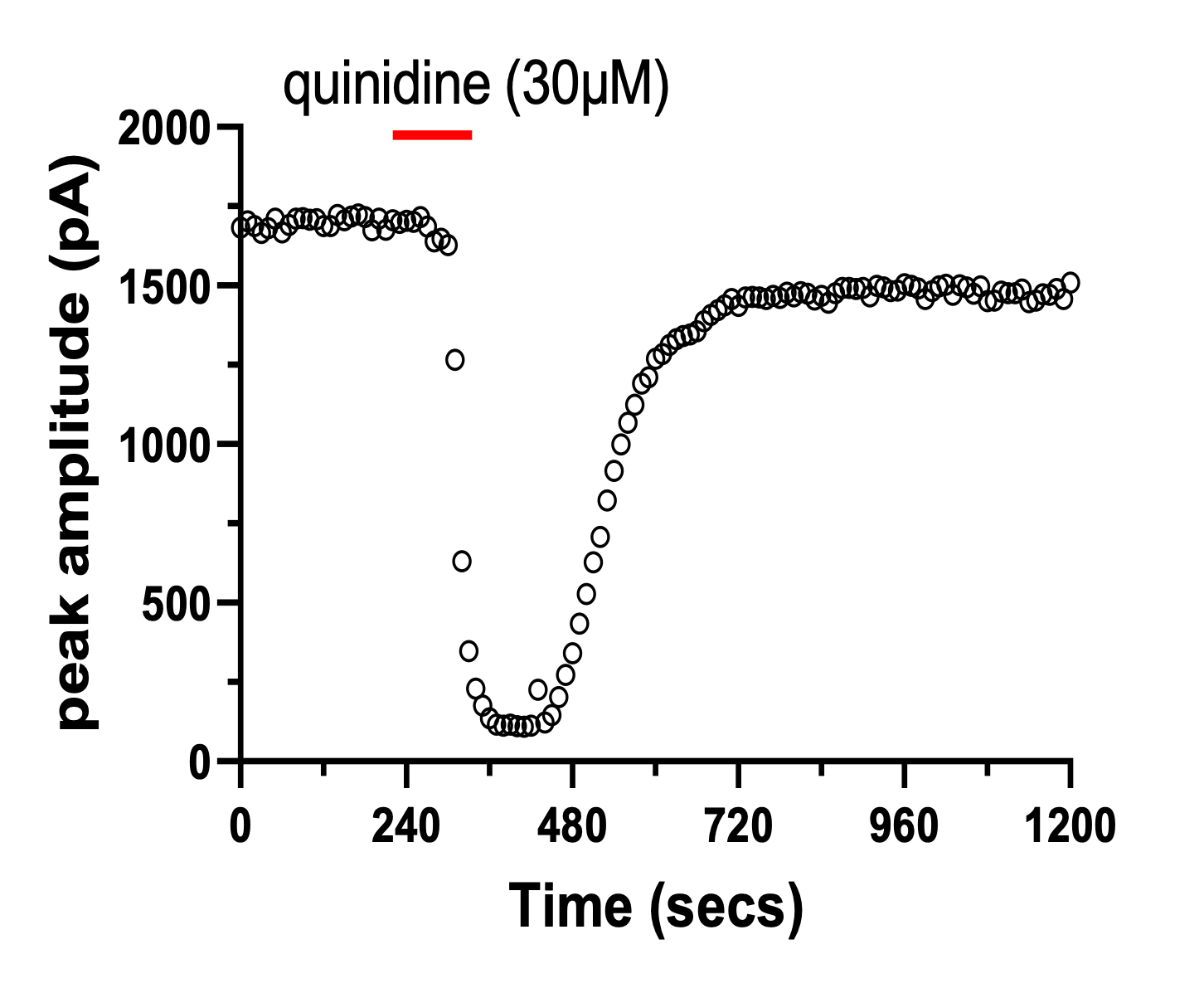
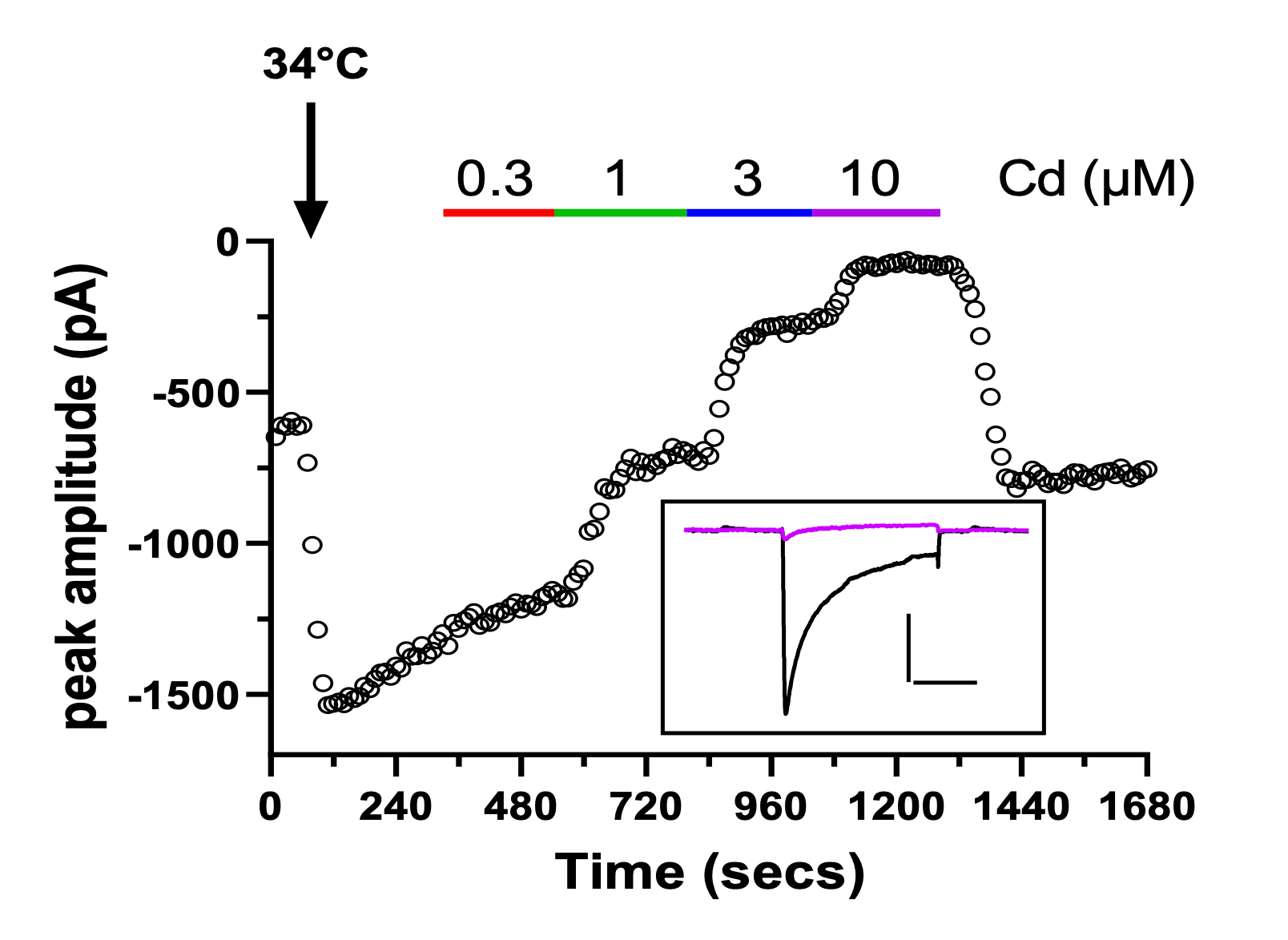
Figure 1 – example time course plots and corresponding traces (insets) recorded from cells stably expressing hERG (A, scale bar 500pA, 500ms) or Cav2.2 (B, scale bar 500pA, 20ms) channels.
A key drawback of MPC is that its low-throughput nature is unconducive to screening large numbers of compounds quickly. Resultantly automated patch-clamp (APC) machines were developed and have largely replaced MPC as the “go-to” technique for ion channel screening. Despite this, MPC affords many advantages, for example:
- Studying the phenotypes of fluorescently tagged transgenes (Figure 2).
- Single channel recordings
- GLP studies
- Assay development
- Assessing the intracellular actions of low-volume compounds.
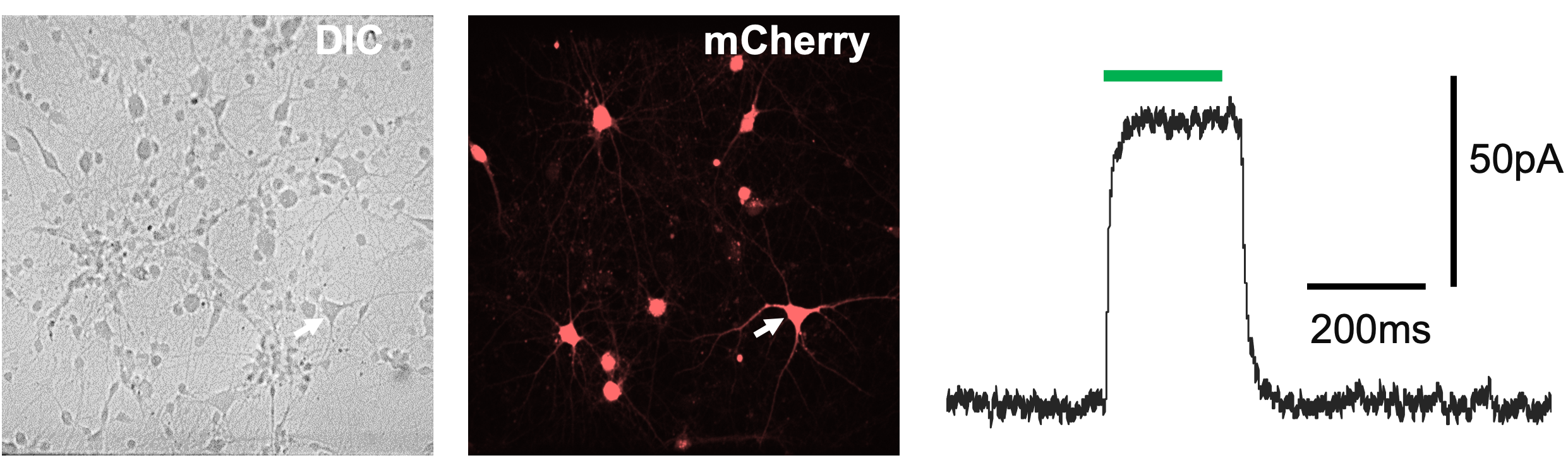
Figure 2– whole-cell MPC recording of fluorescently tagged cell (shown by white arrow)
ApconiX offers both MPC and APC for custom studies tailored to our clients needs.