
Early Ion Channel Screening for Cardiac and Seizure Safety
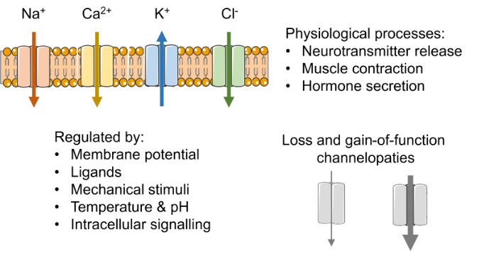
Figure 1: Ion channel function and physiology. Ion channels play a key role in many physiological processes and can be regulated by a diverse range of factors. Abnormal ion channel function, for example loss- or gain-of-function mutations (channelopathies) are associated with a range of conditions.
Ion channels: What are they and why are they important?
Ion channels span the cell membrane and form specialised pores that allow ions such as sodium (Na⁺), potassium (K⁺), calcium (Ca²⁺), and chloride (Cl⁻) to selectively move into or out of cells. Ion channels are usually grouped into families based on the ion they selectively permit to cross the membrane. Each ion-channel family can be further subdivided into members that differ in their regulation, pharmacology, and electrophysiological properties. This complexity makes ion channels well suited to play key roles in physiological processes, including neurotransmitter release, muscle contraction and hormone secretion 1. The physiological importance of ion channels is underlined by conditions caused by abnormal ion channel function (channelopathies) and by their use as drug targets to treat various conditions.
Channelopathies
Channelopathies are diseases caused by abnormal ion channel function and are linked to various conditions 2,3 (Figure 2). They are most commonly caused by genetic mutations in the genes encoding ion channels or their auxiliary β subunits, but can also result from acquired factors such as drugs, toxins, or abnormal electrolyte levels 4,5. The diverse range of channelopathies emphasises the critical role that ion channels play in physiological processes and highlights why they are key targets in drug discovery.
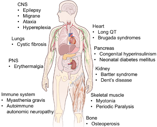
Figure 2: Channelopathies are associated with a range of conditions. Figure illustrates key channelopathies (not a complete list) 2,3.
Ion channels as drug targets
Ion channels are one of the largest target classes, second only to GPCRs by number of approved modulators 6,7, with clinical uses ranging from anaesthesia to disease treatment. Lidocaine, for example, blocks Na⁺ channels, preventing the depolarisation needed for neuronal firing and thereby producing local anaesthesia. Glibenclamide blocks pancreatic β-cell Kₐₜₚ channels, which depolarises the cell and triggers Ca²⁺ influx and insulin release, making it effective in the treatment of type 2 diabetes. Conversely, retigabine enhances neuronal Kᵥ7 ion channel activity to dampen excitability, which underlies its efficacy in epilepsy. Together, these examples illustrate why ion channels are successful drug targets: subtle pharmacological modulation, via blocking or enhancing activity to decrease or increase cell excitability, can achieve a desired therapeutic outcome. Despite their success as drug targets, unintended modulation of ion channels, either through the intended target in the wrong tissue, or through an entirely different off-target ion channel, can result in ion channel-induced toxicity8-10.
Ion channel-induced toxicity
Regardless of a drug’s intended therapeutic use, ion channel-induced toxicity must be considered, as improper modulation of ion channels can disrupt essential biological functions and lead to unacceptable adverse effects. Therefore, detecting ion channel-related toxicity early in drug development is essential to avoid costly late-stage failures.
This article outlines why early ion channel safety screening is essential in drug development, using key examples from cardiac and CNS, and discusses some of the strategies used to avoid ion channel-induced toxicity. Two important safety liabilities addressed by ion channel screening are drug-induced cardiac arrhythmia, assessed through hERG and CiPA strategies, and seizure liability, increasingly assessed using panel-based ion channel screening plus human iPSC-derived neuronal assays.
The role of hERG in cardiac health and drug safety
The hERG (Kᵥ11.1) ion channel is a textbook example of why early ion channel screening is important in drug development 11. Disruption of the hERG current can trigger fatal arrhythmias and has resulted in market withdrawal of several drugs. hERG carries the rapid delayed rectifier current (IKR), a major contributor to action potential repolarisation. When hERG is blocked or mutated, repolarisation is delayed, meaning it takes longer for the cells to return to their resting electrical state. In isolated cells, this delay is seen as an action potential prolongation while clinically it is detected as a prolonged QT interval on an electrocardiogram (ECG). This delay in repolarisation can trigger Torsades de Pointes (TdP), a polymorphic ventricular tachycardia that can degenerate into ventricular fibrillation and sudden death11-13. This mechanism led to several market withdrawals in the late 1990s and early 2000s and prompted the International Council for Harmonisation (ICH) in 2005 to recommend a preclinical in vitro hERG assay 11,14,15. Today, automated patch-clamp platforms screen for hERG liability early in drug development before a programme has absorbed substantial time and cost.

hERG block is not the whole story
Blocking hERG delays ventricular repolarisation and increases the likelihood of Early Afterdepolarisations (EADs). EADs are abnormal additional depolarisations that occur before the action potential has fully repolarised and are considered to be the cellular trigger for TdP. Based on this mechanism alone, any hERG blocker might be assumed to carry proarrhythmic risk.
However, hERG block alone does not determine TdP risk. Compounds can modulate more than one cardiac ion channel, an effect referred to as multiple ion channel effects (MICE). When this includes concurrent block of inward sodium or calcium currents alongside hERG block, the additional block can reduce the likelihood of EADs, lowering proarrhythmic risk compared to hERG block alone 16,17.
As a result, a hERG-only decision framework can both deprioritise compounds with low TdP liability and miss dangerous compounds that act through other key cardiac ion channels 18-20. These limitations of hERG-centric screening prompted the broader multi-channel approach proposed by the Comprehensive in vitro Proarrhythmia Assay (CiPA) initiative, a paradigm that is increasingly used to inform regulatory risk assessment.
Comprehensive in vitro Proarrhythmia Assay
CiPA combines a trifecta of complementary nonclinical assays (Figure 3), with the aim of improving early risk assessment while avoiding unnecessary attrition of compounds that may be safe despite showing hERG activity 20:
- In vitro ion channel screening on several cardiac ion channels.
- An in silico model of a human ventricular action potential which integrates the ion channel data from step 1.
- A human stem-cell-derived cardiomyocyte assay, providing an integrated cellular readout.
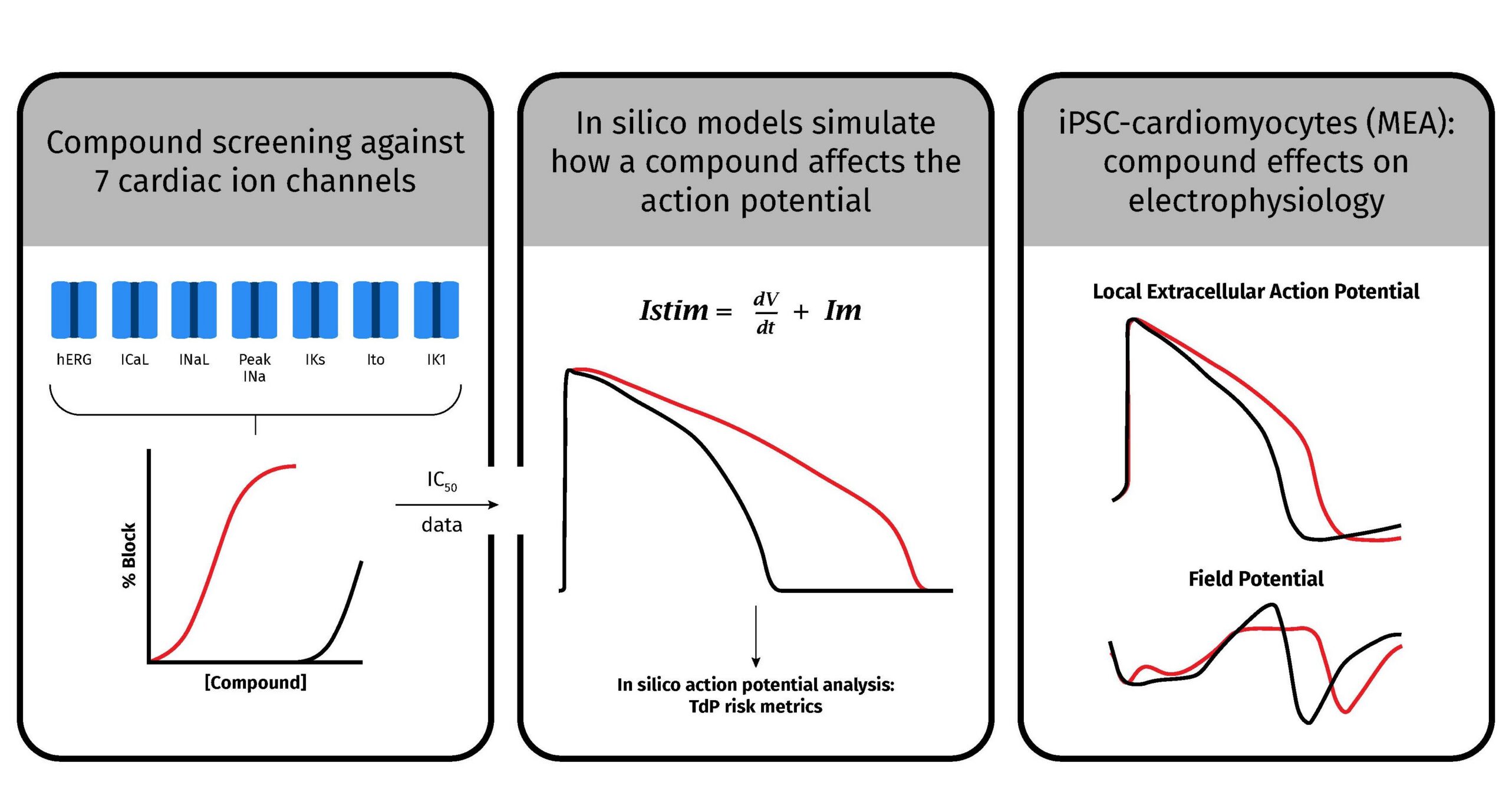
Figure 3: CiPA in vitro framework illustration. Ion channel screening on up to seven cardiac ion channel currents allows the IC₅₀ for a compound to be measured for each channel. The IC₅₀ values are integrated into an in silico model of the ventricular cardiomyocyte from which metrics of proarrhythmic risk are derived. The final in vitro assay uses human iPSC-derived cardiomyocytes which provide a more physiologically complete cellular model capable of detecting missed or unanticipated electrophysiological responses. The red line illustrates a typical hERG blocker: inhibition of hERG at lower concentrations results in prolongation of the in silico action potential, as well as prolongation of the iPSC cardiomyocyte action potential and field potential.
Patch-clamp to detect modulation of multiple cardiac currents
The CiPA ion channel screen includes whole-cell patch-clamp assays on a range of cardiac ion channels, including the outward repolarising currents Kᵥ11.1 (hERG, Iₖᵣ), Kᵥ7.1/KCNE1 (Iₖₛ), Kᵥ4.3 (Iₜₒ), IK1 (Kir2.1) and the inward depolarising Naᵥ1.5 (peak and late) and Caᵥ1.2 currents. These assays are generally run in recombinant expression systems so that each current can be measured in isolation before the integrated effect is considered at the in silico model and cell level.


In silico computer model of the cardiac action potential
In silico models integrate ion channel data to simulate how a compound’s combined effects across multiple channels would alter the human ventricular action potential. From these simulated action potentials, specific metrics can be calculated, which are then used to predict a compound’s TdP risk.

Human induced pluripotent stem-cell-derived cardiomyocytes
Human induced pluripotent stem-cell-derived cardiomyocytes (hiPSC-CMs) capture many key aspects of native cardiomyocyte electrophysiology. Studies using hiPSC-CMs can detect changes in action potential and field potential duration, which are indicators of disturbed electrophysiology 18,20.
Ultimately this trifecta of early preclinical assays can provide a more informative view of proarrhythmic liability than hERG testing alone. When used together, the preclinical CiPA assays can enable early go/no-go decisions in drug development before committing to expensive in vivo studies. More recently, similar approaches have been used to screen for other adverse reactions due to drug-induced modulation of ion channels.
Seizure liability screening
Drug-induced seizures occur frequently in drug development and are usually detected during preclinical animal studies or early clinical trials, leading to significant compound attrition 21. Because so much compound attrition occurs after substantial investment has been made, there is a clear need to detect seizure liability earlier in drug development using in vitro preclinical assays.
Like Long QT (LQT) syndromes, research into genetic and acquired channelopathies identified a role for ion channels in seizure 22. However, unlike drug-induced LQT syndromes, there is not a single ion channel (hERG in LQT) responsible for the majority of drug-induced seizures. Some examples of the ion channels implicated in seizure include sodium (Naᵥ1.1, Naᵥ1.2), calcium (Caᵥ2.1, Caᵥ3.2), and potassium ion channels (Kᵥ1.1, Kᵥ2.1, Kᵥ3.1, Kᵥ4.2, Kᵥ7.2/7.3), as well as ligand-gated ion channels (GABAₐ, Nicotinic α4β2, NMDA1/2A). Further complexity is added by the type of neuron each ion channel is expressed in, the sub-cellular locations of the ion channels within each neuron type and whether the change in the ion channel increases or decreases neuronal excitability 22-25 (Figure 4).
The success of ion channel screening in reducing compound attrition caused by TdP risk 26, 27 led to the successful implementation of CiPA-like in vitro approaches to assess a compound’s seizure liability 23,25,28. First, a panel of neuronal ion channels can be screened using automated patch-clamp assays. Secondly, a hiPSC-neuron-based assay can identify compound-induced alterations in neuronal firing and network bursting associated with seizure liability 25.
Used together, these preclinical assays provide a more complete assessment of seizure risk than either alone. The ion channel panel identifies which specific channels a compound modulates, providing mechanistic insight. The hiPSC-neuron-based assay captures how ion channel-level modulation alters network activity, giving a functional readout that complements the ion channel panel’s mechanistic detail 25. As with CiPA assays in cardiac safety, this combination allows safety teams to make earlier, better-informed go/no-go decisions, helping to reduce CNS-related attrition.
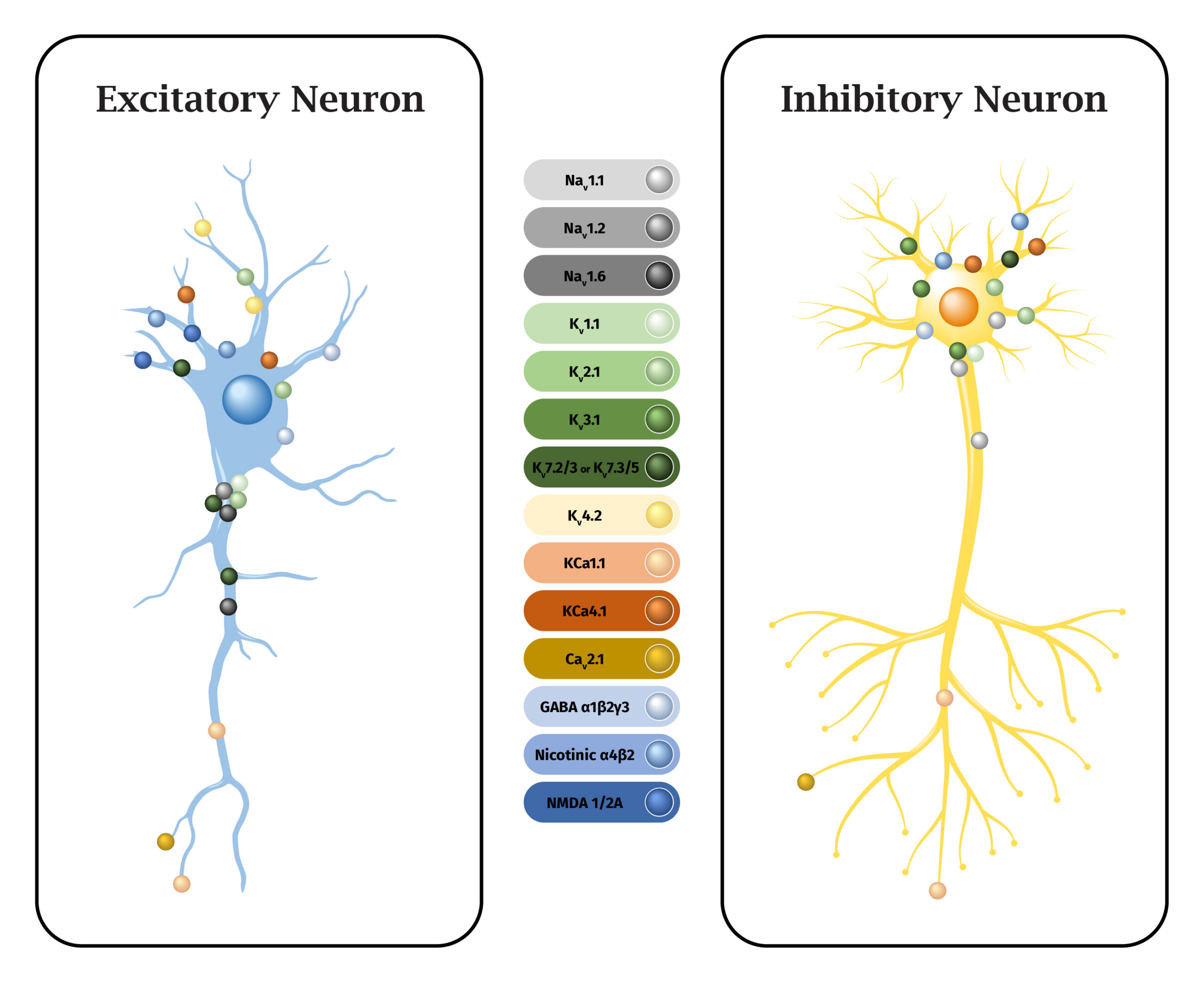
Figure 4: Predominant subcellular locations of key seizure-associated ion channels. Ion channel expression and subcellular localisation vary between excitatory (left) and inhibitory (right) neurons, adding to the functional complexity and differential contribution of each channel to cellular excitability. Channel placements represent the dominant, most-established location(s) reported in the literature and are schematic rather than exhaustive or quantitatively precise 29-53.

ApconiX: Nonclinical safety expertise to biotech, biopharma and academia
ApconiX has an entire division specialising in ion channel target and safety screening. ApconiX combines high-throughput automated patch-clamp systems with manual patch-clamp capability to provide rapid, reliable electrophysiology data for drug development. ApconiX can provide hERG liability data within 4 days, with CiPA and seizure ion channel screening taking an average of 14 days. ApconiX also provides Target Safety Assessments, prepared by experienced toxicologists to identify a target’s key risks by organ/tissue.
Learn more about automated patch clamp
Learn more about ApconiX’s Automated Patch Clamp Services, including available study options and support for ion-channel screening.