
From Voltage Clamp to Automated Patch Clamp
Automated patch clamp developed from decades of electrophysiology research, beginning with voltage clamp and progressing through manual patch clamp to parallel recording systems.
Ion channels are remarkable proteins. Around 400 have been identified in the human genome, and they are expressed in every cell in the human body.
Although ion channels vary greatly in their structure and physiological function, they share a common purpose: allowing ions to pass from one side of the plasma membrane to the other in response to a stimulus.
That stimulus may be a change in membrane potential, in the case of voltage-gated ion channels, or a ligand such as a neurotransmitter, proton or other substance, in the case of ligand gated ion channels.
The development of techniques capable of measuring this activity has transformed scientists’ understanding of ion-channel function.

The foundations of voltage clamp
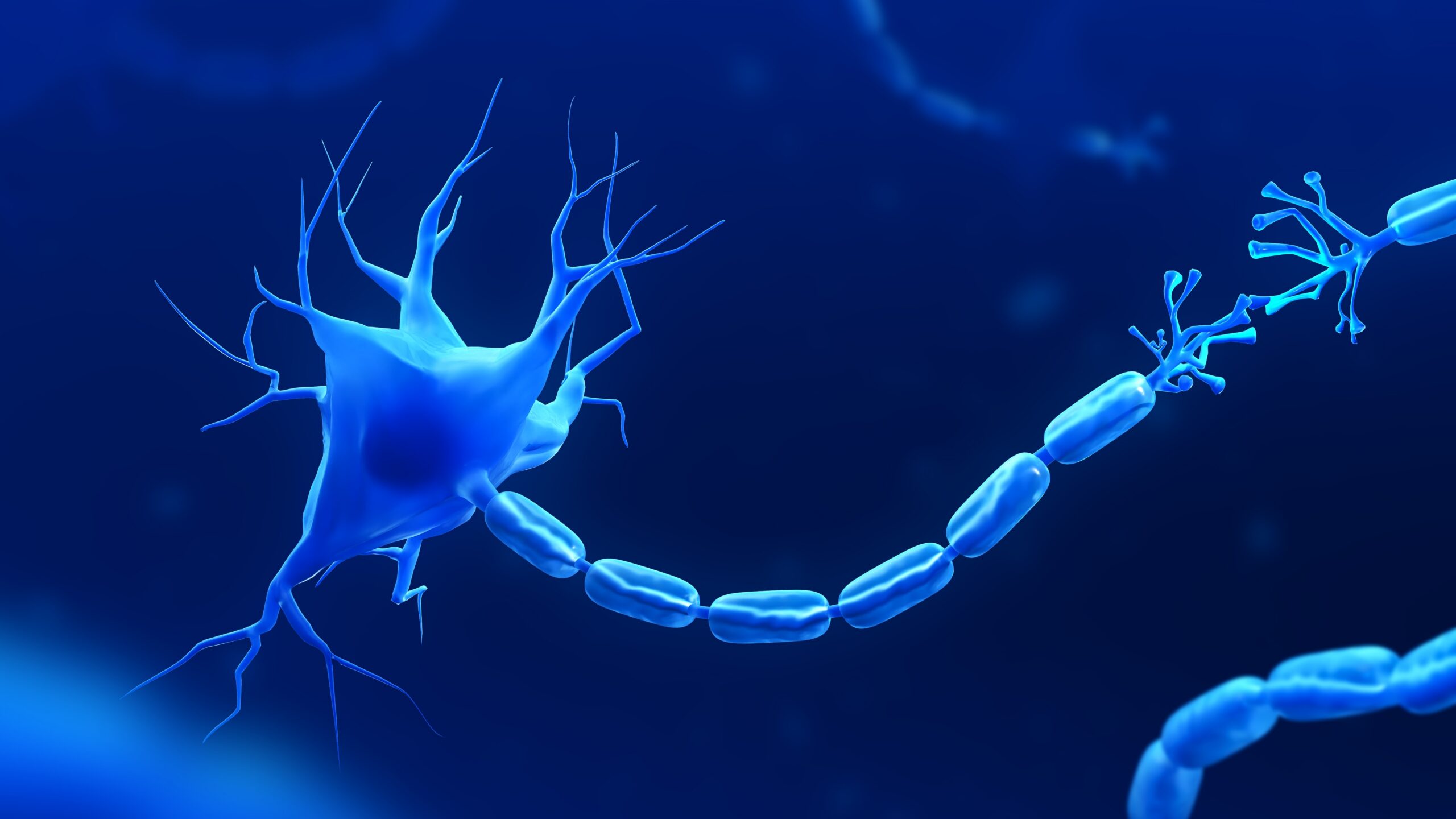
Following the Second World War, Alan Hodgkin and Andrew Huxley studied the squid giant axon and became the first scientists to use the voltage clamp technique.
By impaling the giant axons with electrodes, they were able to record the ionic currents flowing across the axonal membrane without the resulting changes in membrane potential affecting the measurement.
This work was completed before individual ion channels had been identified. Hodgkin and Huxley instead measured ionic “conductances”, allowing them to develop an understanding of the action potential and the movement of sodium and potassium ions across the membrane.
Their work earned them the 1963 Nobel Prize in Physiology or Medicine.
During the 1960s, scientists continued to develop the concept that these conductances resulted from ions passing through pores or channels in the plasma membrane. At the time, the theory was not universally accepted and reportedly faced considerable scientific opposition.
The patch-clamp breakthrough
A major change in the measurement of ion channels came with the development of the patch-clamp technique.
During the mid-1970s, Erwin Neher and Bert Sakmann used a fine glass microelectrode with a microscopic opening. When the electrode was gently pressed against the cell membrane and light suction was applied, it formed a high-resistance seal – known as a gigaseal – between the glass and the membrane.
This isolated a tiny patch of membrane containing one or more ion channels, allowing their activity to be measured directly.
Research published by Sigworth and Neher in 1980 recorded sodium-channel activity in rat muscle. It demonstrated that ion channels fluctuated rapidly between open and closed states and allowed the conductance of individual channels to be measured.
Neher and Sakmann received the 1991 Nobel Prize in Physiology or Medicine for their work.
Since the technique was first developed, several forms of patch clamp have been introduced, including cell-attached, whole cell and perforated-patch recording.
Conventional, or manual, patch clamp remains an important technique for measuring ion-channel activity. Within drug discovery, it may also be used to investigate the effects of compounds on ion channels such as hERG.
Why patch clamp needed to be automated
Although manual patch clamp provides detailed measurements, the technique is slow and requires considerable technical skill.
As pharmaceutical interest in ion channels increased, researchers needed to test much larger numbers of compounds. Conventional patch clamp alone could not provide the throughput required for large drug discovery programmes.
Several alternative technologies were developed to study ion channels without using electrophysiology. These included ligand binding, ion flux and fluorescence based assays.
However, these approaches did not always measure ion channel function directly. A method was therefore required that could increase throughput while retaining the functional measurements provided by patch clamp electrophysiology.

The first automated patch-clamp systems
In a 2002 interview, Kirk Schroeder, the co-developer of IonWorks (an automated patch-clamp system), described the objective of increasing patch-clamp throughput.
The traditional glass pipette, microscope and micromanipulator used in manual patch clamp were replaced by a planar substrate containing hundreds of small holes.
Cells could form high-resistance seals over these holes in parallel. After sealing, a perforating agent allowed the instrument to control, or “clamp”, the membrane potential and record ion channel activity.
An early publication using this technology investigated Kv1.5 and NaV1.3 ion channels. It presented evidence relating to voltage clamp capability, recording success rates, comparative biophysics, pharmacology and assay consistency.
The introduction of automated systems allowed many recordings to be conducted in parallel, substantially increasing the number of compounds that could be investigated.
The development of population patch clamp
An important development followed in 2007 with the introduction of population patch clamp.
In the earlier single hole format, differences between individual cells could create considerable well to well variability. This made it difficult to generate reliable concentration response curves.
Population patch clamp reduced this problem by averaging the signal across multiple cells within an individual well. In the published work, signals from 64 cells were combined.
Averaging the response reduced the effect of cell-to-cell variation and allowed the resulting data to be fitted to generate XC50 (an umbrella term used to describe the concentration of a substance required to achieve 50% of a maximum effect, response, or inhibition in an in vitro (laboratory assay) values.
This represented another important stage in the evolution of automated patch clamp, supporting higher-throughput functional ion channel assessment and more quantitative investigation of compound activity.
Learn more about automated patch clamp
Learn more about ApconiX’s Automated Patch Clamp Services, including available study options and support for ion-channel screening.