
Cardiac MEA Assays for Drug Discovery
We offer comprehensive cardiac microelectrode (MEA) assays for cardiac safety screening (including action potential measurement and proarrhythmia detection) using human induced pluripotent stem cell (hiPSC) derived cardiomyocytes. As one of the key pillars of the Comprehensive in vitro Proarrhythmia Assay (CiPA), cardiomyocyte MEA screening enables you to directly measure electrical activity and ion channel function and forms part of our integrated approach to cardiac safety.
What is cardiac MEA screening?
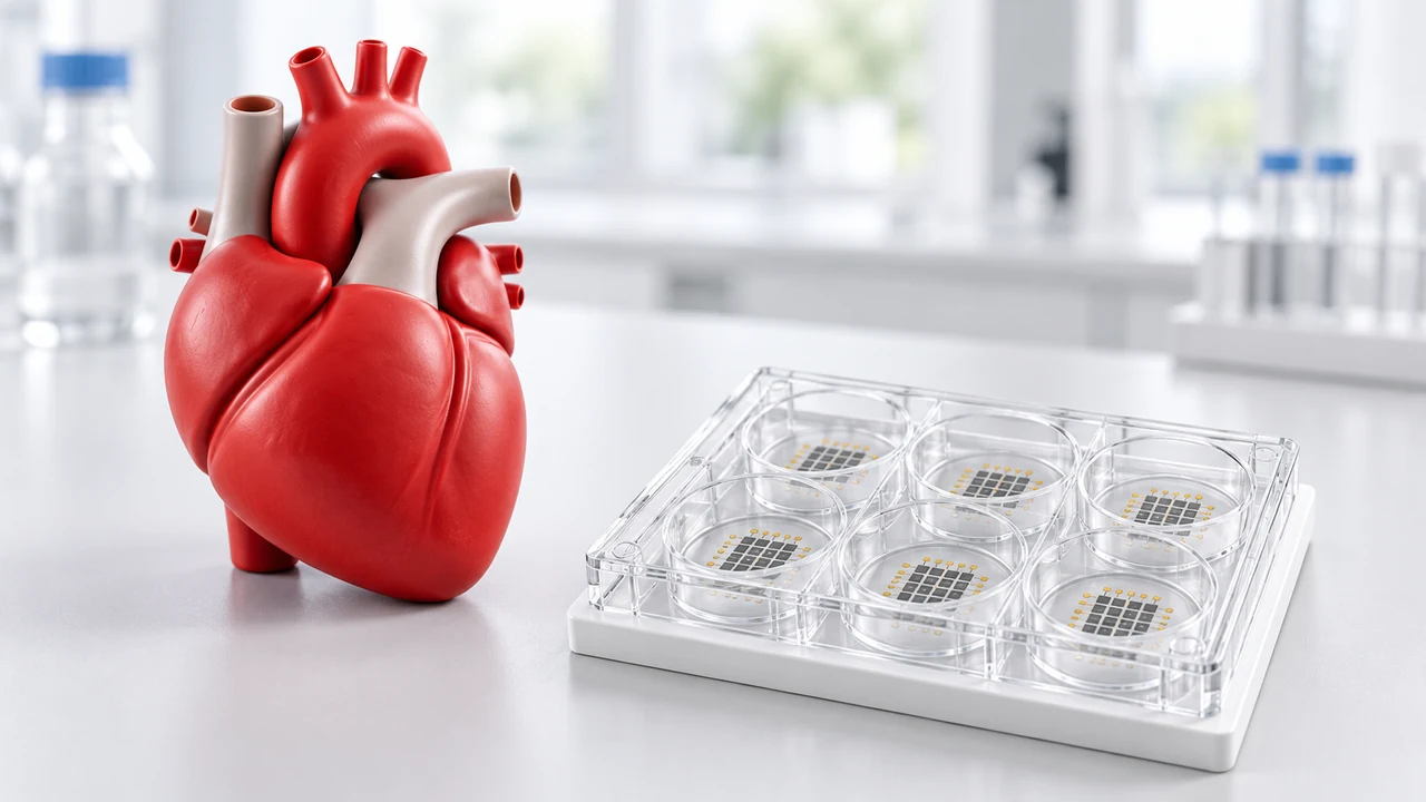
In drug discovery, we use cardiac MEA screening to evaluate how a drug compound impacts cardiac electrophysiology.
The drug of interest is administered to a network of hiPSC cardiomyocytes that are grown on a specialised grid of microscopic electrical sensors. These sensors can detect electrical signals coming from the cardiomyocytes in response to the drug in a highly sensitive, noninvasive way.
Cardiac MEA screening can show:
- conduction velocity – record how fast electrical signals travel across cells. Used for detecting risk for heart blocks or circuit short overs
- Propagation pattern – map the path of the electrical signal across the cell plate. Used for detecting chaotic beating patterns
- Beat rate and rhythm regularity – record the speed and pattern of cardiomyocyte network pulsing. Used for arrythmia detection (Torsades de Pointes).
- Field potential duration – measure how long it takes for the cardiac cell network to repolarise after a beat. This repolarisation related measurement helps to assess QT prolongation risk.


Why use hiPSC derived cardiomyocytes?
hiPSC derived cardiomyocytes offer direct modelling of human biology, including in their expression of human ion channels and proteins. Since hiPSC derived cardiomyocytes form a network (mimicking cardiac tissue) you can monitor whole tissue reactions to a drug, such as coordinated cell pulsing, rather than looking at response in a single, isolated cell.
It’s important to be aware that hiPSC cardiomyocytes don’t always behave like mature, adult cells and tend to more closely resemble foetal cardiomyocytes in nature. Additionally, it’s worth noting that although hiPSCs cardiomyocytes form a network which models heart tissue beautifully, they are still isolated from other organs and biological systems unlike in vivo.
What can ApconiX measure?
Our cardiomyocyte screening service can provide a range of readout metrics using FPD and Local Extracellular Action Potential (LEAP) assays in addition to impedance modules using our in-lab Maestro Pro MEA Platform (Axion Biosystems):
| Readout metric | Indication | Assay module type |
|---|---|---|
| Beat rate | Chronotropic effects, e.g. bradycardia and tachycardia. | Cardiac module (LEAP assay) |
| Field potential duration (FPD) | Detects delays in repolarisation. | Cardiac module (FPD) |
| Sodium spike amplitude and slope | A measure of Vmax which can be used as an indicator of a sodium channel block. | Cardiac module (FPD) |
| Action potential duration (APD) at 30%, 50% and 90% repolarisation | Measures the time taken for cells to repolarise after depolarisation. Prolonged APD can indicate inhibition of the hERG channel. | Cardiac module (LEAP assay) |
| Rise time | The amount of time for the cell to completely depolarise from its resting potential. | Cardiac module (LEAP assay) |
| Triangulation ratio | Calculates a change of shape of the action potential which indicates underlying electrical instability. | Cardiac module (LEAP assay) |
| Early Afterdepolarisations (EADs) | Abnormal, additional depolarisations that occur during the plateau or repolarisation phases that can initiate arrhythmias. | Cardiac module (LEAP assay) |
| Contraction amplitude / vector | Identifies changes in cardiomyocyte contractility (inotropy). | Cardiac module (impedance) |
| Impedance baseline / viability | Measures cell attachment and monolayer integrity. Differentiates functional cardiotoxicity from general cytotoxicity. | Impedance module |
What do I need to provide for the Cardiac MEA assay?
We require:
- 5-10 mg of your drug compound
- Your compound’s molecular weight
- The number of dosing concentrations you will need
To help us best tailor our cardiac MEA assay to your specific needs, we also find having any available information regarding the solubility of your compound and its Cmax very helpful.



What will I receive and when?
We will provide you with a highly detailed cardiac MEA report, including:
- Details of the experiments performed, who carried out the work and a comprehensive summary of experimental methods
- Representative plots showing cardiac cell activity at each test concentration and across each electrode in each MEA well for easy visual comparison of drug induced effects on cell activity
- Graphical presentation of all replicates to show the degree of variability in the dataset and other Quality Control (QC) information
- Summarised results of the MEA analysis presented in a clear, colour coded table
- An invitation to discuss your results with our experts
Our Cardiac MEA assay usually takes 3-4 weeks. However, it’s important to bear in mind that different factors can affect the turnaround for our Cardiac MEA service. Of course, our experts are always on hand to discuss how we can best meet the needs of your project and its specific timelines.
When to use cardiac MEA screening
Our Cardiac MEA service can be used to support both drug discovery and development for:
- Compound comparison: directly compare the phenotypic response to different drug candidates at the cellular network level
- Liability investigation: assess key cardiac events, such as block of hERG, proarrhythmia, and cytotoxicity as part of your cardiotoxicity screening
- Mechanistic follow up: use MEA to assess whole cell response to drug administration and combine with other available methods, such as our ion channel panel, to gain better mechanistic insight into drug induced cardiac liability
- Acute / time dependent effects: evaluate cardiac liability in response to administration of an acute dose or repeated dosing to mimic a clinical regimen
Our expert team can work with you help you determine how our Cardiac MEA service might best fit the needs of your particular project.



Study options and design
At ApconiX, we understand that every drug discovery project is unique. We can therefore offer a wide range of cardiac MEA study options and designs to suit your needs, including:
- Drug exposure
- Drug concentrations
- Study controls
- Repeats
- Pacing
How does cardiac MEA fit into a wider cardiac safety context?
Since MEA assessment bridges the gap between high throughput molecular screening and later stage in vivo testing, it serves as a critical component to our multitiered Cardiac Safety service, which includes:



These tools, along with our Cardiac MEA assay, form key pillars of the CiPA framework. Our Cardiac MEA assessment will build a more complete picture of potential cardiac safety issues to support better decision making.
Example Study: Investigation of potential cardiotoxicity in hiPSC-cardiomyocytes using the Maestro Pro MEA assay system (Axion Biosystems)
In this study, we show how our Cardiac MEA service can be used to build a picture of compound cardiac liability before costly in vivo work begins:
Real time compound effect on APD (Figure 1a) and changes in morphology are identified using LEAP. This allows for the clear visualisation of arrhythmias, for example EADs, (Figure 1b).
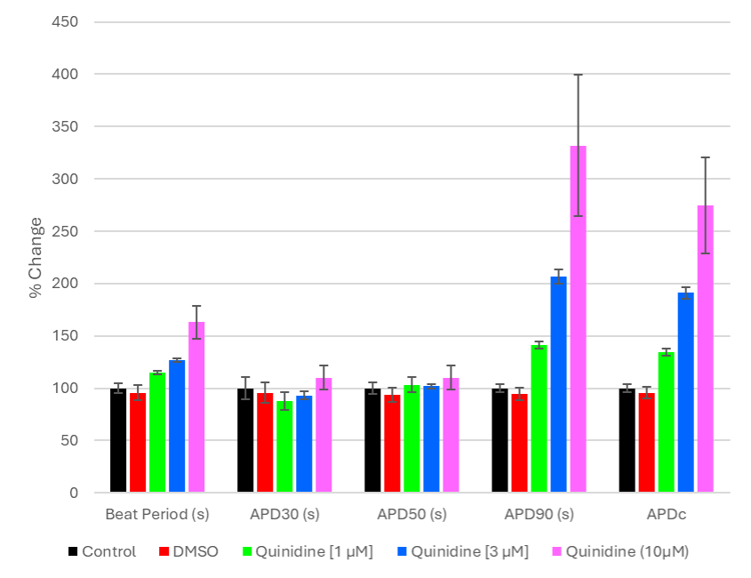
Figure 1a. LEAP metrics measured under normal conditions and in the presence of increasing concentrations of Quinidine.
Quinidine addition (Na+ and K+ channel blocker) results in the prolongation of repolarisation giving rise to an increase in beat period and action potential duration.
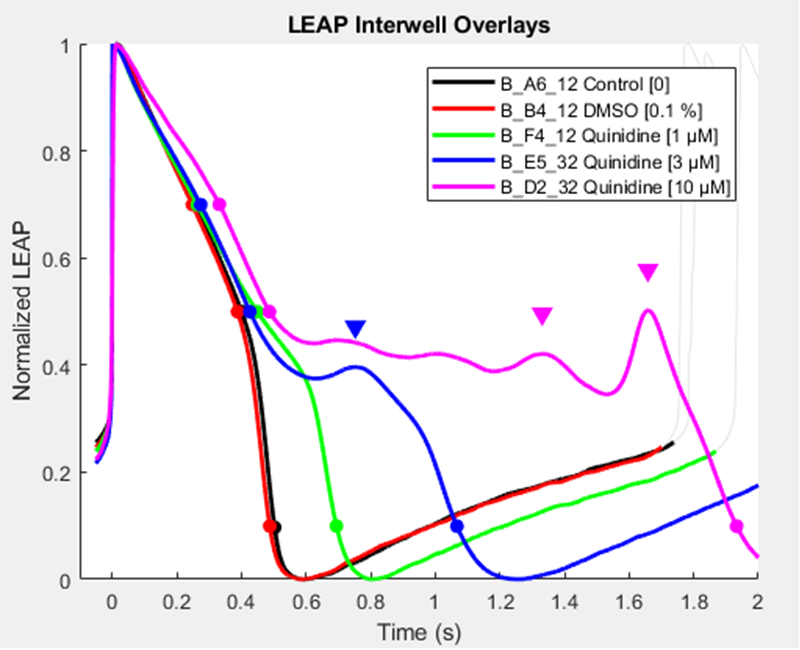
Figure 1b. LEAP measurements observed under normal conditions and in the presence of Quinidine
Quinidine at low dose causes a prolongation of repolarization, as the dose increases EAD’s are observed.